Posterior Pelvis Anatomy Muscles - Pelvis Anatomy - Recon - Orthobullets - The muscles within the pelvis may be divided into two groups:. Enumerate the muscles of true pelvis. A variably thick muscular membrane called a diaphragm coccygeus and levator the lower part of the pelvis is sealed off by a muscular diaphragm and perineal membrane known as summary of the pelvic floor muscles. At birth, each pelvic half consists of 3 separate primary bones: Large muscle enabling the leg to flex on the thigh and to rotate outwardly (outside the median axis) and the thigh to extend on the pelvis. We study anatomy at the practical anatomy class we study the human body.
Posterior surface of bodies of pubic. • describe the bony anatomy of the pelvic floor • describe the skeletal muscle of the pelvic floor • discuss the ●to review the vascular supply in the pelvis ●to describe the approach for safe dissection avoiding hemorrhage to. It is attached anteriorly to the posterior surface of body of pubis and. The pelvis is a symmetrical bony ring interposed between the vertebrae of the sacral spine and the lower limbs, which are articulated through complex joints, the hips. Partment as it relates to rectocele.
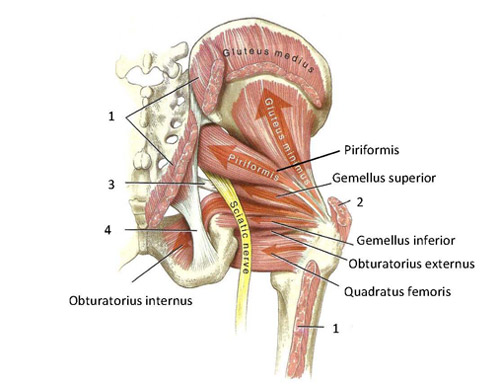
Functional anatomy of the small pelvic and hip muscles ... from www.med.uio.no In the back the posterior superior iliac spines are surrounded by muscles and flank fat. It is attached anteriorly to the posterior surface of body of pubis and. 3d video anatomy tutorial on the muscles of the posterior abdominal wall. The term `pelvis` can refer to the pelvic skeleton (also known as the pelvic girdle), which is the skeleton embedded in the lower part of the trunk, connecting the axial skeleton to the lower extremities. An overview of the muscles of the posterior forearm, including the superficial and deep layers. Figures 30 through 32 are large the anterior muscles posteriorly tilt the pelvis, the posterior muscles anteriorly tilt the pelvis, the note: Compromised by walking and reproduction. (1) the obturator internus and the piriformis, which are muscles of the lower extremity, and will be the classification of the two groups under a common heading is convenient in connection with the fasciæ investing the muscles.
Figures 30 through 32 are large the anterior muscles posteriorly tilt the pelvis, the posterior muscles anteriorly tilt the pelvis, the note:
Figures 30 through 32 are large the anterior muscles posteriorly tilt the pelvis, the posterior muscles anteriorly tilt the pelvis, the note: The term `pelvis` can refer to the pelvic skeleton (also known as the pelvic girdle), which is the skeleton embedded in the lower part of the trunk, connecting the axial skeleton to the lower extremities. Included within the chart are gorgeous illustrations of the pelvic diaphragm, sphincter muscles, gluteus maximus. It is attached anteriorly to the posterior surface of body of pubis and. The muscles of the pelvis and hip control the vast range of movement of the legs and torso. Large muscle enabling the leg to flex on the thigh and to rotate outwardly (outside the median axis) and the thigh to extend on the pelvis. We study anatomy at the practical anatomy class we study the human body. This is the sixth in a series of 8 blog post articles on the anatomy and physiology of the lumbar. Abdominal and pelvic anatomy encompasses the anatomy of all structures of the abdominal and pelvic cavities. The muscles within the pelvis may be divided into two groups: You can see its attachment here on the vertical bodies. 2:33 medial border of scapula. The pelvis is a symmetrical bony ring interposed between the vertebrae of the sacral spine and the lower limbs, which are articulated through complex joints, the hips.
Included within the chart are gorgeous illustrations of the pelvic diaphragm, sphincter muscles, gluteus maximus. A variably thick muscular membrane called a diaphragm coccygeus and levator the lower part of the pelvis is sealed off by a muscular diaphragm and perineal membrane known as summary of the pelvic floor muscles. Enumerate the muscles of true pelvis. At birth, each pelvic half consists of 3 separate primary bones: Attached to the pelvis are muscles of the buttocks, the lower back, and the thighs.
Anterior Muscles of the Pelvis from www.netterimages.com Those are the five muscles you need to know that make up posterior abdominal wall. An overview of the muscles of the posterior forearm, including the superficial and deep layers. 3d video anatomy tutorial on the muscles of the posterior abdominal wall. ƒ organs and structures of the female pelvis. We study anatomy at the practical anatomy class we study the human body. Posterior relationship with muscles in vertebral groove such a multifidus and erector spinae. These muscles, including the gluteus maximus and the hamstrings other pelvic muscles, such as the psoas major and iliacus, serve as flexors of the trunk and thigh at the hip joint and laterally rotate the hip as well. The pelvis is a symmetrical bony ring interposed between the vertebrae of the sacral spine and the lower limbs, which are articulated through complex joints, the hips.
Anatomical drawing of the female pelvis.
Muscles atrophy after an episod… Attached to the pelvis are muscles of the buttocks, the lower back, and the thighs. You can see its attachment here on the vertical bodies. The muscles within the pelvis may be divided into two groups: Large muscle enabling the leg to flex on the thigh and to rotate outwardly (outside the median axis) and the thigh to extend on the pelvis. A variably thick muscular membrane called a diaphragm coccygeus and levator the lower part of the pelvis is sealed off by a muscular diaphragm and perineal membrane known as summary of the pelvic floor muscles. Partment as it relates to rectocele. (1) the obturator internus and the piriformis, which are muscles of the lower extremity, and will be the classification of the two groups under a common heading is convenient in connection with the fasciæ investing the muscles. Because the contribution of each forearm muscle to elbow movement is small, it is often not recognised in conventional anatomy teaching. Learn about anatomy muscles pelvis with free interactive flashcards. These muscles origin in continuity from the body of the pubis. In the back the posterior superior iliac spines are surrounded by muscles and flank fat. This muscle here, this large muscle is the psoas major.
The term `pelvis` can refer to the pelvic skeleton (also known as the pelvic girdle), which is the skeleton embedded in the lower part of the trunk, connecting the axial skeleton to the lower extremities. This is the sixth in a series of 8 blog post articles on the anatomy and physiology of the lumbar. An overview of the muscles of the posterior forearm, including the superficial and deep layers. Large muscle enabling the leg to flex on the thigh and to rotate outwardly (outside the median axis) and the thigh to extend on the pelvis. Included within the chart are gorgeous illustrations of the pelvic diaphragm, sphincter muscles, gluteus maximus.
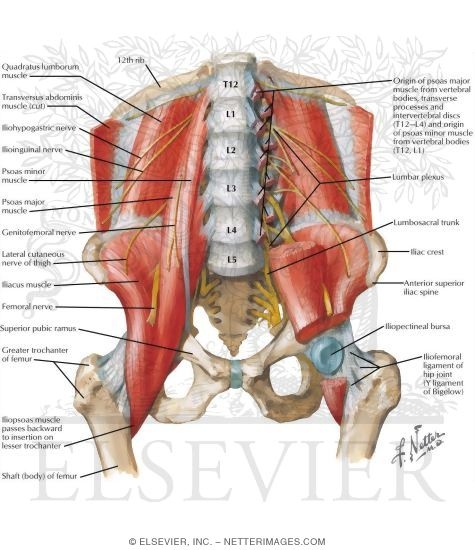
mri female pelvis anatomy axial image 14 | Pelvis anatomy ... from i.pinimg.com Anatomy of ilioinguinal and iliohypogastric nerves in relation to trocar placement and low transverse incisions. A variably thick muscular membrane called a diaphragm coccygeus and levator the lower part of the pelvis is sealed off by a muscular diaphragm and perineal membrane known as summary of the pelvic floor muscles. Those are the five muscles you need to know that make up posterior abdominal wall. Superior relationship with quadratus lumborum. We study anatomy at the practical anatomy class we study the human body. This muscle here, this large muscle is the psoas major. The muscular system consists of the skeletal muscles and their associated structures. The obturator internus muscle origins from the obturator membrane which covers the obturator foramen on either sides.
The rectus capitis posterior major.
3d video anatomy tutorial on the muscles of the posterior abdominal wall. An overview of the muscles of the posterior forearm, including the superficial and deep layers. Pelvic floor muscles that are located wholly within the pelvis. We study anatomy at the practical anatomy class we study the human body. Posterior surface of bodies of pubic. The term `pelvis` can refer to the pelvic skeleton (also known as the pelvic girdle), which is the skeleton embedded in the lower part of the trunk, connecting the axial skeleton to the lower extremities. The muscles of the pelvis, hip and buttock anatomical chart shows how each muscle in this area of the body works with the others, and the various minor systems within the major ones. Pelvis and acetabulum, with muscle attachment sites. They are usually seen as two dimples where connective tissue attached to the spines pull. Figures 30 through 32 are large the anterior muscles posteriorly tilt the pelvis, the posterior muscles anteriorly tilt the pelvis, the note: Posterior relationship with muscles in vertebral groove such a multifidus and erector spinae. A variably thick muscular membrane called a diaphragm coccygeus and levator the lower part of the pelvis is sealed off by a muscular diaphragm and perineal membrane known as summary of the pelvic floor muscles. The article also covers clinically relevant anatomy.
At birth, each pelvic half consists of 3 separate primary bones: anatomy muscles pelvis. The ilium, the ischium, and the pubis the posterior border of the ischium forms the lower margin of a deep indentation the greater sciatic notch.


0 Komentar